




 JDVS Home



Abstract
This study described the detection and molecular characterization of Bovine viral diarrhea virus (BVDV) field strains in two mummified bovine fetuses and a persistently infected (PI) heifer from a vaccinated closed Holstein dairy cattle herd located in Teixeira Soares, in the southcentral region of Paraná State, southern Brazil. Tissue fragments from the mummified fetuses (brain, lung, liver, small intestine, and placenta) and three serial blood samples from all the animals in the herd were submitted to reverse transcription (RT)-PCR assay for the partial amplification of the 5’-untranslated region (5’UTR) and N- terminal auto protease (Npro) gene of the Orthopestivirus genome. Additionally, the fetal tissue samples were tested for other reproductive pathogens. A total of 44 serum samples from heifers (n = 15) and cows (n = 29) were evaluated for BVDV antibodies using the virus neutralization test. Amplicons with high DNA quantification from the fetal tissue and blood serum of the PI heifer in the 5’UTR and Npro RT-PCR assays were sequenced. BVDV1b was detected in all fetal tissues and the blood serum of the PI heifer, with 100% nucleotide identity. All samples from both mummified fetuses tested negative for Bovine alphaherpesvirus 1, Brucella spp., Histophilus somni, Leptospira spp., Mycoplasma bovis, Mycoplasma bovigenitalium, Neospora caninum, and Ureaplasma diversum. The PI heifer was BVDVseronegative in the virus neutralization test, while 97.7% (43/44) of other pregnant and non-pregnant cows in the herd were BVDV-seropositive. This study documents the first case of BVDV infection in a mummified fetus from a PI pregnant heifer.
Keywords:Bovine; Orthopestivirus bovis; Bovine viral diarrhea virus; Reproductive disease; Abortion
Abbreviations:BVDV: Bovine Viral Diarrhea Virus; 5’UTR: 5’-Untranslated Region; RT: Reverse Transcription; BoAHV1: Bovine Alphaherpesvirus 1; FTAI: Fixed-Time Artificial Insemination; TI: Transiently Infected; DEPC: Diethylpyrocarbonate; VN: Virus Neutralization
Introduction
Among the distinct clinical challenges in bovine reproduction, such as heat repetitions at regular or irregular intervals, abortion, fetal maceration, stillbirth, and neonatal mortality, fetal mummification has sporadic occurrence [1,2]. Due to its low frequency, the etiology of fetal mummification is often neglected; however, it is recognized as a multi-etiological condition. Bovine viral diarrhea, leptospirosis, neosporosis, mycotoxins, umbilical cord compression or torsion, uterine torsion, defective placentation, genetic anomalies, abnormal hormonal profiles, and chromosomal abnormalities have all been associated with this unusual condition [1,3]. Bovine viral diarrhea virus (BVDV) is an endemic viral pathogen affecting dairy and beef cattle herds worldwide. BVDV is a major infectious agent primarily associated with reproductive and respiratory disorders, and it is responsible for significant economic losses due to its detrimental impact on cattle productivity [4,5]. Orthopestiviruses are currently classified into 19 species, including Orthopestivirus bovis (BVDV1), Orthopestivirus tauri (BVDV2), and Orthopestivirus brazilense (BVDV3 or Hobi-like Orthopestivirus), which are the species known to infect bovine hosts [6].
Additionally, at least 24 subgenotypes of BVDV1 (BVDV1a to BVDV1x) have been described [7], along with five subgenotypes of BVDV2 (BVDV2a to BVDV2e) [8], and five subgenotypes of BVDV3 (BVDV3a to BVDV3e) [9]. In Brazil, BVDV infection is considered endemic in both beef and dairy cattle herds across all geographic regions [10]. The virus is associated with reproductive [11], respiratory [12], enteric [13], mucosal [14], and neurological disorders [15]. Although BVDV was first linked to bovine fetal mummification in 1973 [16], few spontaneous cases have been documented since [3,17,18]. To date, molecular characterization of the BVDV strain involved in bovine fetal mummification has been performed only once, identifying a wild-type BVDV1a strain in an unvaccinated small dairy cattle herd [17]. This study reports the detection and molecular characterization of BVDV field strains in two mummified bovine fetuses from a closed, BVDV-vaccinated, dairy cattle herd with a history of reproductive failures. Notably, this is the first documented case of BVDV infection in a mummified fetus from a persistently infected (PI) dam.
Materials and methods
Herd, sanitary profile, and reproductive history
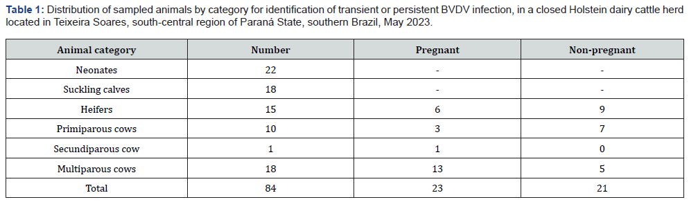
Two cases of fetal mummification occurred within a ninemonth interval between them in a closed Holstein dairy cattle herd located in Teixeira Soares, in the south-central region of Paraná State, southern Brazil. The distribution of the 84 females (calves, heifers, and cows) of the herd by animal category is shown in Table 1. The dairy herd had an average milk production of 23 L per cow per day. The lactating cows were maintained on Cynodon spp. cv. Tifton 85 pastures, supplemented with corn silage and commercial concentrate. Access to commercial mineral supplements and water was ad libitum. Sanitary management practices to prevent infectious reproductive diseases included a biannual vaccination program for cows, conducted in March and September, using polyvalent vaccines containing inactivated Bovine alphaherpesvirus 1 (BoAHV1), BVDV1a and 2a, and Leptospira spp. (serovars Pomona, Canicola, Icterohaemorrhagiae, Hardjo, and Grippotyphosa). The herd was officially certified free of brucellosis and tuberculosis. Reproduction was performed exclusively using fixed-time artificial insemination (FTAI). One year before the mummification events, reproductive failures were documented but not investigated, including a low conception rate, averaging six inseminations per confirmed pregnancy, early embryonic mortality, and 12 abortions occurring between the third and eighth months of pregnancy. During the evaluation period, there were no reports of neonatal or suckling calf deaths.
Fetal mummification cases
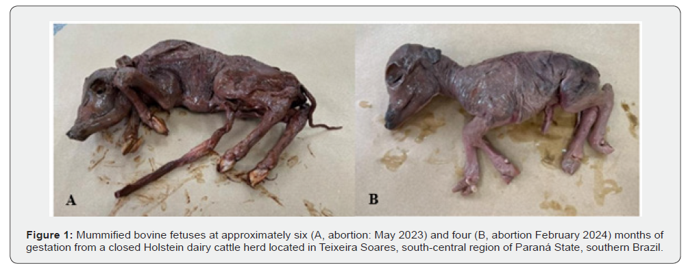
Case #1: In May 2023, a pregnant Holstein heifer exhibited estrus eight months after FTAI. During the genital examination, the veterinarian detected a mummified fetus in the vaginal canal, which was removed by hand. The mummified fetus was male, measuring 45 cm in length, corresponding to approximately 6 months of gestation (Figure 1A). Case #2: In February 2024, a secundiparous Holstein cow exhibited estrus six months after FTAI. During the genital examination, fetal death was confirmed by the veterinarian. Two days later, the cow aborted a mummified female fetus measuring 31 cm in length, corresponding to approximately 4 months of gestation (Figure 1B). The fetuses and blood serum samples from their progenitors were immediately refrigerated and sent for laboratory analysis.
Biological samples
Tissue fragments from the brain, lung, liver, small intestine, and placenta were collected from mummified fetuses and stored at -80 ºC until analysis. To monitor BVDV circulation in the dairy cattle herd and to diagnose transiently infected (TI) and PI animals, three serial blood samplings were conducted from May to July 2023 at 30-day intervals (Table 1). All 23 pregnant animals had their newborn calves monitored for BVDV presence in blood serum after birth. Calves RT-PCR BVDV-positive in the first test would be shown again 30 days later. Blood samples without anticoagulant were also collected from all pregnant and non-pregnant females in the herd to obtain serum for the identification of anti-BVDV antibodies. The serum was separated by centrifugation at 3,000 x g for 5 min and stored in sterile microtubes at -80 ºC until further processing.
Molecular investigation
All tissue samples from the two mummified fetuses were homogenized using a TissueLyser LT® (QIAGEN, Hilden, Germany) for 5 min with 0.01M phosphate-buffered saline (PBS) in 10% suspension (w/v), and subsequently centrifuged at 2,000 x g for 5 min. Aliquots of 500 μL of the supernatants were incubated at 56 ºC for 30 min with sodium dodecyl sulfate and proteinase K, at final concentrations of 1% (v/v) and 0.2 mg/mL, respectively. Nucleic acids were extracted using a combination of phenol/chloroform/ isoamyl alcohol (25:24:1) and silica/guanidine isothiocyanate techniques [19,20]. The extracted nucleic acids were eluted in 50 μL of ultrapure sterile water treated with diethylpyrocarbonate (DEPC) (Invitrogen, Carlsbad, CA, USA) and stored at -80 ºC until use. For blood serum samples, 500 μL of each sample was processed according to the method previously described [19-28].
The nucleic acid was eluted in 50 μL of DEPC-treated water (Invitrogen, Carlsbad, CA, USA) and immediately stored at -80 ºC until use. Nucleic acids extracted from the fetal tissue samples were submitted to molecular assays to investigate etiologic agents known to cause reproductive disorders in cattle, as described in Table 2. Additionally, nucleic acids extracted from serum samples were subject to an RT-PCR assay to amplify the partial sequence of the 5′ UTR and Npro gene of BVDV. As the negative control for all nucleic acid extractions, DEPC-treated water was used. As positive controls, cell culture-adapted (Madin-Darby bovine kidney, MDBK) BVDV Singer strain, MDBK-adapted BoAHV1 Los Angeles strain, as well as DNA of Neospora caninum, Leptospira interorgan serovar Hardjo, Histophilus somni, Brucella abortus, Mycoplasma bovis, Mycoplasma bovigenitalium, and Ureaplasma diversum derived from previous investigation were used [17]. The molecular assay products were analyzed by electrophoresis on 2% agarose gels prepared in Tris-boric acid-EDTA buffer, pH 8.4 (89 mM Tris; 89 mM boric acid; 2 mM EDTA), containing ethidium bromide (0.5 μg/mL).
Electrophoresis was performed at a constant voltage of 100 V for 40 min. Following electrophoresis, the agarose gels were visualized under ultraviolet light and photo documented. The amplicons obtained by molecular techniques were purified using the Quick Gel Extraction and PCR Purification Combo Kit (Invitrogen Life Technologies, Carlsbad, CA, USA). They were quantified with a Qubit® Fluorometer (Invitrogen Life Technologies, Eugene, OR, USA), and sequenced in an ABI3500 Genetic Analyzer (Applied Biosystems®, Foster City, CA, USA) using the same forward and reverse primers used in the previous assay with the BigDye® Terminator v3.1 Cycle Sequencing Kit (Applied Biosystems, Foster City, CA, USA). Sequence quality analysis was performed using PHRED software, and contigs assembled using CAP3 software (http://asparagin.cenargen.embrapa.br/phph/). The nucleotide (nt) sequences were compared with sequences deposited in GenBank using BLAST software (http://blast. ncbi.nlm.nih.gov/Blast.cgi). Phylogenetic trees based on the nt sequences were constructed using the Neighbor-joining method with the Kimura two-parameter model in MEGA software version 10.2.6 [29]. Bootstrapping was statistically supported with 1,000 replicates. The nt sequence identity matrices were performed in BioEdit software version 7.2.5 [30].
Serological test
Serum samples collected at the occasion of case #1 (15 heifers and 29 cows - Table 1) were evaluated for the presence of antibodies against BVDV using the virus neutralization (VN) test, performed according to the Manual of Diagnostic Tests and Vaccines for Terrestrial Animals of the World Organization for Animal Health [31]. Serum samples were inactivated at 56 ºC for 30 min and then diluted from 1:8 to 1:512. For the VN test, the virus used was the cell culture (MDBK) adapted NADL (BVDV1a) strain. The titer of the viral strain used in the VN test was standardized to 100 TCID50. Virus and serum dilutions were incubated for 1 h at 37 ºC in a 5% CO2 atmosphere. Afterward, 50 μL of 3 × 10⁵ MDBK cells/mL was added. The interpretation of the results was performed after 72 h of incubation at 37 ºC. The neutralizing titer considered for each serum sample was the reciprocal of the highest dilution able to neutralize viral replication. Titers ≥8 were considered BVDV-positive. MDBK cells used in the VN test were cultured in Dulbecco’s Modified Eagle’s Medium supplemented with L-glutamine, antibiotics, fungicide, and 10% BVDV-free fetal bovine serum, and regularly tested for BVDV RNA by RT-PCR.
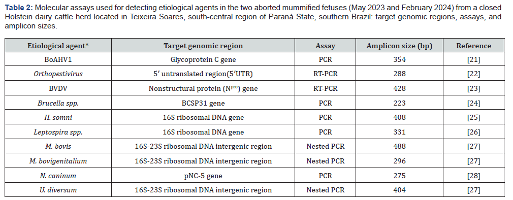
*BoAHV1 (Bovine alphaherpesvirus 1), BVDV (Bovine viral diarrhea virus), H. somni (Histophilus somni), M. bovis (Mycoplasma bovis), M. bovigenitalium (Mycoplasma bovigenitalium), N. caninum (Neospora caninum), U. diversum (Ureaplasma diversum).
Results
Amplicons of the expected size for the 5’UTR and the Npro gene of Orthopestivirus/BVDV were detected in all tissue fragments analyzed of both mummified fetuses. All biological samples from both mummified fetuses were negative in the molecular assays for the other etiological agents evaluated as differential diagnoses. In the three RT-PCR tests performed to detect the 5’UTR of Orthopestivirus and the Npro gene of BVDV in the serum of the 84 animals, only one heifer was identified as having a persistent infection (PI). No other animal showed transient BVDV infection. It is important to highlight that the previously mentioned PI heifer was the mother of the mummified fetus case #1 of this study. Amplicons from the 5’UTR of Orthopestivirus and the Npro gene obtained from the placenta, liver, and small intestine of both mummified fetuses, as well as from the blood serum of the PI heifer, were sequenced to confirm the specificity of the amplified products and to characterize the species and sub genotype. All sequenced amplicons from the mummified fetuses and the PI heifer serum belonged to Orthopestivirus bovis sub genotype 1b. As the nt sequences obtained from different tissues of the same mummified fetuses were identical, only one representative sequence per fetus was used for phylogenetic analysis [32].
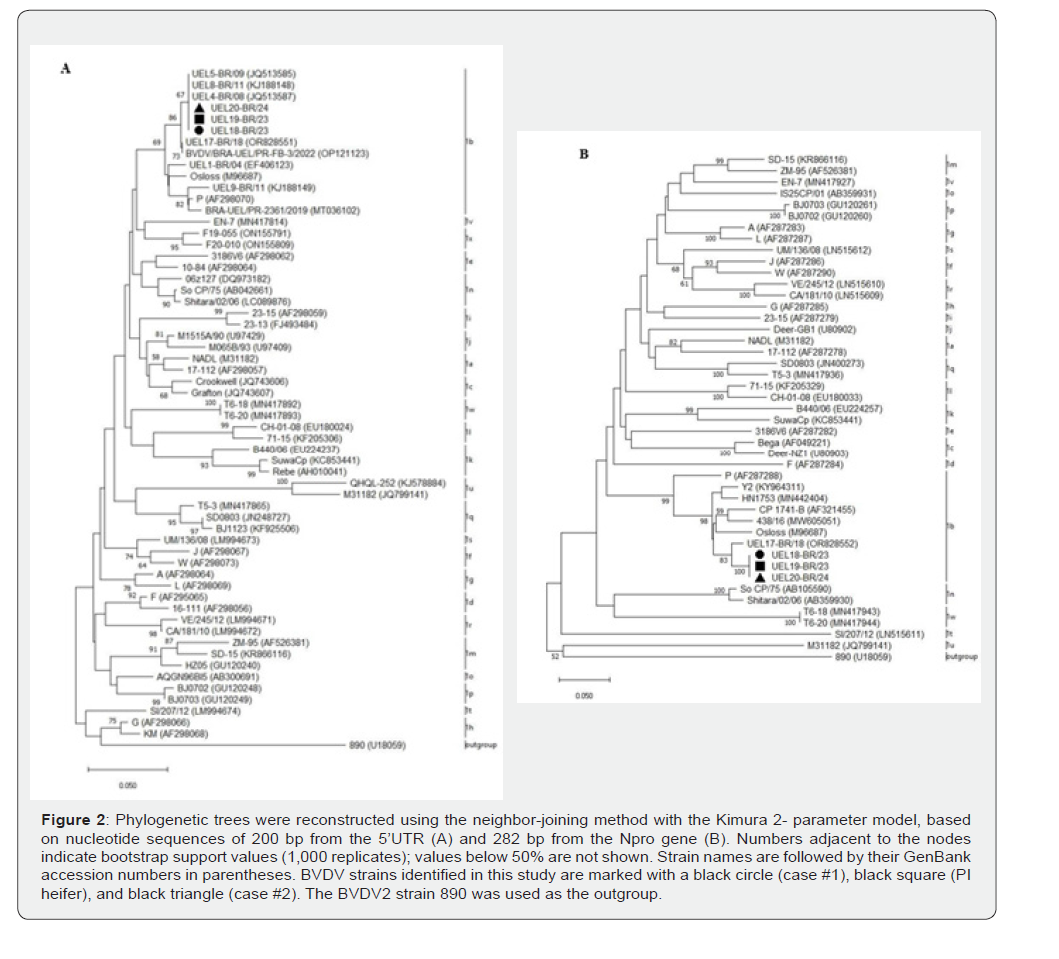
In the 5’UTR genomic analysis, the three BVDV1b sequences obtained in this study from mummified fetuses #1 (UEL18-BR/23), #2 (UEL20-BR/24), and from the PI heifer (UEL19-BR/23) showed 100% of nt identity among them and 94.3 to 100% of nt identity with other BVDV1b strains previously described. When compared to the BVDV1b prototype strains P and Osloss, the sequences exhibited 94.8 and 95.3% of nt identity, respectively. Notably, they shared 100% nt identity with the Brazilian BVDV1b strains UEL4- BR/08 (GenBank accession number: JQ513587) and UEL5- BR/09 (GenBank accession number: JQ513585) [11]. Additionally, nt sequence identity analysis of the Npro amplicons revealed that the three BVDV1b sequences described herein shared 100% of nt identity with each other and 87.9 to 97.1% of nt identity with other previously described BVDV1b strains [32,33]. The highest nt identity (97.1%) was observed with the Brazilian strain UEL17- BR/18 (GenBank accession number: OR828552) [33]. When compared to the
BVDV1b prototype strains P and Osloss, the sequences showed 87.9 and 93.6% of nt identity, respectively.
Phylogenetic analyses based on the 5’UTR genomic (Figure 2A) and the Npro gene (Figure 2B) showed that the three wildtypes BVDV strains identified in this study clustered with the prototype BVDV1b strains, as well as with other BVDV1b strains previously reported in Brazil and other countries. The nucleotide sequences described in the present study have been deposited in the GenBank database under accession numbers: 5’ UTR genomic – strains: UEL18-BR/23 (PX486805), UEL19-BR/23 (PX486806), and UEL20-BR/24 (PX486807); Npro gene – strains: UEL18-BR/23 (PX484914), UEL19-BR/23 (PX484915), and UEL20-BR/24 (PX484916). BVDV-VN antibodies were detected in 97.7% (43/44) of the serum samples analyzed. Among the 43 seropositive animals, 55.8% (n = 24) showed intermediate (64-256) titers and 44.2% (n = 19) had high (≥512) titers of neutralizing antibodies against BVDV. Only the heifer, progenitor of case #1, tested negative. The secundiparous cow, progenitor of case #2, showed a seroconversion rate with a titer of ≥512.
Discussion
This report describes two cases of fetal mummification that occurred within nine months in a closed, BVDV-vaccinated dairy cattle herd with reproductive failure. Although more commonly observed in pluriparous and polytocous species such as swine, fetal mummification remains a rare event in cattle [3]. Fetal mummification in cattle is a multifactorial reproductive disorder associated with both infectious and non-infectious causes [3]. Among infectious agents, both experimental and natural BVDV infections in pregnant cows have been associated with the occurrence of fetal mummification [3,16-18]. In the two cases described here, in addition to screening and the exclusion of the other eight pathogens previously associated with reproductive diseases in bovines, molecular analysis detected only BVDV RNA. In cases of fetal mummification, molecular methods are essential for determining the etiology, as clinical and gross examination alone cannot differentiate the causative agent. However, the prolonged interval between fetal death, dehydration, and expulsion may compromise molecular diagnosis due to the degradation of nucleic acids and proteins [3,17]. In both cases described, highquality amplicons were obtained, allowing partial sequencing of the 5’ UTR and the Npro gene of BVDV from placental, hepatic, and intestinal tissues of the mummified fetuses.
In Brazil, two other isolated cases of fetal mummification associated with BVDV infection have been reported. In the first case, BVDV was detected in a spontaneously aborted bovine fetus at eight months of gestation, approximately 45 cm in length, likely dead since six months of gestation, from a non-BVDV- vaccinated Girolando dairy cattle herd in the northern region of Paraná state. Amplicons were obtained by RT-PCR for partial amplification of the BVDV 5′ UTR in kidney and spleen samples, and the Npro gene in the kidney sample [17]. In a second case, fetal mummification occurred in a Jersey herd vaccinated against both BVDV1 and BVDV2 in Santa Catarina state. The fetus tested positive by RTPCR for the 5´UTR in spleen and thymus samples, although the Npro gene was not analyzed. Additionally, the genome of Neospora caninum was detected by PCR using Np21 and Np6 primers in the brain sample of the mummified fetus [18]. Phylogenetic trees and identity matrices analyses of the nt sequences from the 5´UTR and the Npro gene amplicons from the two cases of fetal mummification and the PI progenitor heifer allowed the identification of the BVDV sub genotype circulating in the dairy cattle herd. All three field strains were classified as BVDV1b, exhibiting high nt identity with previously described BVDV1b field strains. The first molecular characterization of a BVDV field strain associated with bovine fetal mummification worldwide was the report of BVDV1a in Brazil [17]. In contrast, a case described previously did not allow determination of the BVDV sub genotype due to the low quality of the amplicon obtained [18]. The studies highlight the circulation of two distinct BVDV1 subgenotypes associated with fetal mummification in Paraná state: BVDV1b, identified in the current report, and BVDV1a, previously characterized [17].
The BVDV1b strains detected in the mummified fetuses and PI heifer are similar to other BVDV1b strains that have previously been reported to circulate in Brazilian cattle herds. They have been detected in fetal bovine serum [34,35], cattle serum [11,36,37], aborted fetuses [38], animals exhibiting respiratory and digestive clinical diseases [38,39], associated with bovine neonatal diarrhea [13], in a fatal acute BVDV outbreak in beef cattle [14], and in PI animals [11,40]. However, this is the first report of BVDV1b in mummified fetuses. The molecular epidemiology of BVDV in Southern Brazil is further elucidated by the high genetic proximity between the strains identified in this report and regional isolates. Specifically, the infecting BVDV1b strain showed the highest (97.1%) nt identity with the Brazilian strain UEL17-BR/18 (GenBank: OR828552) characterized from an outbreak involving PI heifer calves in another BVDV-vaccinated dairy herd in Paraná State [33]. The close phylogenetic relationship between these strains highlights the consistent circulation and persistence of sub genotype 1b in the same region. Although BVDV1a is the most frequently detected sub genotype in Brazilian cattle herds, followed by BVDV2b [10], BVDV1b remains the predominant sub genotype across the Americas, Asia, and Europe [41].
The VN test performed on the entire herd at the time of case #1 confirmed BVDV infection in the herd. Among the cows tested, 54.5% (n = 24) exhibited intermediate neutralizing antibody titers (64-256), while 43.2% (n = 19) showed high (≥512) titers. Although the herd was regularly vaccinated with a polyvalent inactivated vaccine containing BVDV1a and BVDV2a strains, commercial vaccines rarely induce antibody titers higher than 256 [42]. Therefore, the high titers identified suggest the circulation of BVDV field strains. Despite the BVDV1a and BVDV2a vaccination, cases of fetal mummification were associated with BVDV1b infection. This observation suggests that the immune response elicited by the vaccine strains was insufficient to confer effective cross-protective immunity against the heterologous BVDV1b field strain. This apparent vaccine failure may have been triggered by the presence in the herd of a BVDV1b-infected PI heifer, which, due to continuous viral shedding, increases the infection pressure. In case #1 described in this report, the pregnant heifer was BVDV-PI and seronegative for BVDV by the VN test. In case #2, the pregnant cow tested negative for BVDV by RT-PCR but was seropositive, with a high (≥512) antibody titer indicating a previous transient infection. This suggests that the cow experienced a transient BVDV infection that resulted in fetal loss followed by mummification.
Conclusion
Molecular detection and characterization confirmed BVDV1b as the etiological agent associated with fetal mummification and the PI case observed in a BVDV-vaccinated dairy cattle herd in Paraná State, Brazil. To the best of our knowledge, this is the first report of BVDV-associated fetal mummification involving a PI progenitor, and it also describes two occurrences of this rare phenomenon on the same farm. These findings highlight that regular vaccination alone may be insufficient to prevent and control BVDV infection, which is aggravated by the presence of a PI animal.
Acknowledgements
The authors thank the following Brazilian Institutes for financial support: National Institute of Science and Technology of Dairy Production Chain (INCT-LEITE II - Grant number 408.896.2024/8), Araucaria Foundation (FAP-PR), National Council for Scientific and Technological Development (CNPq), Coordination of Superior Level Staff Improvement (CAPES), and Studies and Projects Funding (FINEP). Alfieri, A.A., and Alfieri, A.F are recipients of CNPq fellowships.
References
- Brownlie J (1990) The pathogenesis of bovine virus diarrhea virus infections. Rev Sci Tech 9: 43-59.
- Grooms DL (2004) Reproductive consequences of infection with bovine viral diarrhea virus. Vet Clin North Am Food Anim Pract 20: 5-19.
- Lefebvre RC (2015) Fetal mummification in the major domestic species: current perspectives on causes and management. Vet Med 8(6): 233-244.
- Pinior B, Firth CL, Richter V, Lebl K, Trauffler M, et al. (2017) A systematic review of financial and economic assessments of bovine viral diarrhea virus (BVDV) prevention and mitigation activities worldwide. Prev Vet Med 137: 77-92.
- Scharnböck B, Roch FF, Richter V, Funke C, Firth CL, et al. (2018) A meta-analysis of bovine viral diarrhea virus (BVDV) prevalences in the global cattle population. Sci Rep 8(1): 14420.
- International Committee on Taxonomy of Viruses - ICTV (2025) Master Species List (MSL41).
- Rivas J, Hasanaj A, Deblon C, Gisbert P, Garigliany MM (2022) Genetic diversity of bovine viral diarrhea virus in cattle in France between 2018 and 2020. Front Vet Sci 9: 1028866.
- Oliveira PSB, Silva Júnior JVJ, Weiblen R, Flores EF (2022) A new (old) bovine viral diarrhea virus 2 subtype: BVDV-2e. Arch Virol 167(12): 2545-2553.
- Kalaiyarasu S, Mishra N, Jayalakshmi K, Selvaraj P, Sudhakar SB, et al. (2022) Molecular characterization of recent HoBi-like pestivirus isolates from cattle showing mucosal disease-like signs in India reveals emergence of a novel genetic lineage. Transbound Emerg Dis 69(2): 308-326.
- Flores EF, Cargnelutti JF, Monteiro FL, Bauermann FV, Ridpath JF, Weiblen R (2018) A genetic profile of bovine pestiviruses circulating in Brazil (1998-2018). Anim Health Res Rev 19(2): 134-141.
- Otonel RA, Alfieri AF, Dezen S, Lunardi M, Headley SA (2014) The diversity of BVDV subgenotypes in a vaccinated dairy cattle herd in Brazil. Trop Anim Health Prod 46: 87-92.
- Fritzen JTT, Yasumitsu CY, Silva IV, Lorenzetti E, Alfieri AF (2024a) Respiratory illness in young and adult cattle caused by bovine viral diarrhea virus sub genotype 2b in singular and mixed bacterial infection in a BVDV-vaccinated dairy herd. Braz J Microbiol 55(4): 4139-4146.
- Dall Agnol AM, Lorenzetti E, Leme RA, Ladeia WA, Mainardi RM, et al. (2021) Severe outbreak of bovine neonatal diarrhea in a dairy calf rearing unit with multifactorial etiology. Braz J Microbiol 52(4): 2547-2553.
- Lunardi M, Headley SA, Lisbôa JA, Amude AM, Alfieri AA (2008) Outbreak of acute bovine viral diarrhea in Brazilian beef cattle: clinicopathological findings and molecular characterization of a wild-type BVDV strain subtype 1b. Res Vet Sci 85(3): 599-604.
- Barreto JVP, Lorenzetti E, Fritzen JTT, Jardim AM, Oliveira TES (2022) Congenital neurological disease associated with HoBi-like pestivirus infection in a newborn dairy calf from Brazil. Front Vet Sci 9: 852965.
- Scott FW, Kahrs RF, De Lahunte A, Brown TT, McEntee K (1973) Virus induced congenital anomalies of the bovine fetus. I. Cerebellar degeneration (hypoplasia), ocular lesions and fetal mummification following experimental infection with bovine viral diarrhea-mucosal disease virus. Cornell Vet 63(4): 536-560.
- Fritzen JTT, Morettin AB, Lorenzetti E, Alfieri AF, Alfieri AA (2021) Bovine viral diarrhea virus sub genotype 1a in a mummified fetus from a Brazilian dairy cattle herd. J Vet Diagn Invest 33(5): 966- 968.
- Marian L, Withoeft JA, Esser M, Dal Molin SR, Hamckmeier D, et al. (2024) Uncommon bovine viral diarrhea virus subtype 1e associated with abortions in cattle in southern Brazil. J Vet Diagn Invest 36(1): 115-119.
- Boom R, Sol CJ, Salimans MM, Jansen CL, Wertheim-van Dillen PM (1990) Rapid and simple method for purification of nucleic acids. J Clin Microbiol 28: 495-503.
- Alfieri AA, Parazzi ME, Takiuchi E, Médici KC, Alfieri AF (2006) Frequency of group A rotavirus in diarrheic calves in Brazilian cattle herds, 1998-2002. Trop Anim Health Prod 38: 521-526.
- Claus MP, Alfieri AF, Folgueras-Flatschart AV, Wosiacki SR, Médici KC (2005) Rapid detection and differentiation of bovine herpesvirus 1 and 5 glycoprotein C gene in clinical specimens by multiplex- PCR. J Virol Methods 128: 183-188.
- Vilcek S, Herring AJ, Herring JA, Nettleton PF, Lowings JP (1994) Pestiviruses isolated from pigs, cattle and sheep can be allocated into at least three genogroups using polymerase chain reaction and restriction endonuclease analysis. Arch Virol 136(3-4): 309-323.
- Vilcek S, Paton DJ, Durkovic B, Strojny L, Ibata G, et al. (2001) Bovine viral diarrhea virus genotype 1 can be separated into at least eleven genetic groups. Arch Virol 146(1): 99-115.
- Baily GG, Krahn JB, Drasar BS, Stoker NG (1992) Detection of Brucella melitensis and Brucella abortus by DNA amplification. J Trop Med Hyg 95(4): 271-275.
- Angen O, Ahrens P, Tegtmeier C (1998) Development of a PCR test for identification of Haemophilus somnus in pure and mixed cultures. Vet Microbiol 63: 39-48.
- Marianelli C, Tarantino M, Astarita S, Martucciello A, Capuano F (2007) Molecular detection of Leptospira species in aborted fetuses of water buffalo. Vet Rec 161(9): 310-312.
- Voltarelli DC, De Alcântara BK, Lunardi M, Alfieri AF, De Arruda Leme R (2018) A nested-PCR strategy for molecular diagnosis of mollicutes in uncultured biological samples from cows with vulvovaginitis. Anim Reprod Sci 188: 137-143.
- Baszler TV, Gay LJ, Long MT, Mathison BA (1999) Detection by PCR of Neospora caninum in fetal tissues from spontaneous bovine abortions. J Clin Microbiol 37(12): 4059-4064.
- Kumar S, Stecher G, Li M, Knyaz C, Tamura K (2018) MEGAX: Molecular evolutionary genetics analysis across computing platforms. Mol Biol Evol 35: 1547-1549.
- Hall TA (1999) Bio Edit: a user-friendly biological sequence alignment editor and analysis program for Windows 95/98/NT. Nucleic Acids Symp Ser 41: 95-98.
- World Organization for Animal Health - WOAH (2023) Manual of Diagnostic Tests and Vaccines for Terrestrial Animals.
- Gallina L, Koch MC, Gentile A, Treglia I, Bombardi C, et al. (2021) Bovine viral diarrhea virus 1b infection associated with congenital tremor and hypomyelination in Holstein calves. Vet Microbiol 256: 109047.
- Fritzen JTT, Zucoloto NZ, Lorenzetti E, Alfieri AF, Alfieri AA (2024b) Outbreak of persistently infected heifer calves with bovine viral diarrhea virus subgenotypes 1b and 1d in a BVDV-vaccinated open dairy herd. Acta Trop 254: 107198.
- Silveira S, Weber MN, Mósena AC, Da Silva MS, Streck AF, et al. (2017) Genetic diversity of Brazilian bovine pestiviruses detected between 1995 and 2014. Transbound Emerg Dis 64(2): 613-623.
- Monteiro FL, Cargnelutti JF, Braunig P, Folgueras-Flatschart AV, Santos NC, et al. (2018) Detection and genetic identification of pestiviruses in Brazilian lots of fetal bovine serum collected from 2006 to 2014. Braz J Vet Res 38: 387-392.
- Weber MN, Silveira S, Machado G, Groff FH, Mósena AC, et al. (2014) High frequency of bovine viral diarrhea virus type 2 in Southern Brazil. Virus Res 13(191): 117-124.
- Monteiro FL, Martins B, Cargnelutti JF, Noll JG, Weiblen R (2019) Genetic identification of pestiviruses from beef cattle in southern Brazil. Braz J Microbiol 50: 557-563.
- Cortez A, Heinemann MB, De Castro AMMG, Soares RM, Pinto AMV, et al. (2006) Genetic characterization of Brazilian bovine viral diarrhea virus isolates by partial nucleotide sequencing of the 5'-UTR region. Braz J Vet Res 26(4): 211-216.
- Bianchi E, Martins M, Weiblen R, Flores EF (2011) Genotypic and antigenic profile of bovine viral diarrhea virus isolates from Rio Grande do Sul, Brazil (2000-2010). Braz J Vet Res 31(8): 649-655.
- Dias FC, Médici KC, De Sousa RLM, Alfieri AA, Samara SI (2012) Genetic characterization of bovine viral diarrhea virus detected in persistently infected cattle in Southern Minas Gerais, Brazil. Braz J Vet Res Anim Sci 49(6): 452-458.
- Yeşilbağ K, Alpay G, Becher P (2017) Variability and global distribution of subgenotypes of bovine viral diarrhea virus. Viruses 9(6): 128.
- Potgieter LN (1995) Immunology of bovine viral diarrhea virus. Vet Clin North Am Food Anim Pract 11(3): 501-520.

























