Ultra-High Resolution Electrocardiography
Zaichenko KV, Gurevich BS** and Kordyukova AA
Institute for Analytical Instrumentation, Russian Academy of Sciences, 31-33 Ivana Chernych Str., St. Petersburg, Russia
Submission: April 11, 2024; Published: April 19, 2024
*Corresponding author: Boris S Gurevich, Institute for Analytical Instrumentation, Russian Academy of Sciences, 31-33 Ivana Chernych Str., St. Petersburg, 198095, Russia
How to cite this article: Zaichenko KV, Gurevich BS, Kordyukova AA. Ultra-High Resolution Electrocardiography. 2024; 19(2): 556010 DOI: 10.19080/JOCCT.2024.19.556010
Abstract
Ultra-high resolution electrocardiography (UHR ECG) is a new author’s method of recording and processing of electrocardiac signals (ECS) with ultra-high resolution. Only this method provides extraction of all useful information from the analyzed ECS. It is based on studies of their fine structure with the ultimate goal of early diagnosis of cardiac pathologies, primarily ischemic heart disease (IHD). The relevance of such studies is due to the fact that, according to the World Health Organization (WHO), ischemic heart disease is one of the most common causes of mortality in the population. In 2022, 15.5 million deaths from cardiovascular pathologies were recorded. In addition, early diagnosis of cardiac pathologies based on the detection of their new electrocardiographic markers and diagnostically significant signs will be one of the advantages of using electrocardiography in routine screening of cardiovascular diseases, since ECG is much cheaper than other methods for early diagnosis of IHD - MRI, CT and PET.
Keywords: Cardiac pathology; Electrocardiac signal; Fine structure; Methods of searching for signs of cardiac pathology
Abbreviations: UHR ECG: Ultra-High Resolution Electrocardiography; ECS: Electrocardiac Signal; IHD: Ischemic Heart Disease; MRI: Magnetic Resonance Imaging; CT: Computer Tomography; PET: Positron Emission Tomography; CVS: Cardiovascular System
Introduction
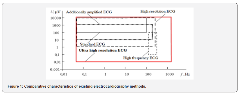
Development of the new UHR ECG method is carried out according to the fundamental scientific concept adopted by the team of one of the leading scientific schools of the Russian Federation, “Radioelectronic and information means of assessment of physiological parameters of living systems” headed by Professor, Dr. K.V. Zaichenko - extraction of the maximum amount of information from the obtained UHR ECG. The scientific team of this school initiated the development and subsequent elaboration of the UHR ECG method based on the application of the latest achievements of radio-electronic, radar and information technologies. Due to this, one of the main ideas of UHR ECG method is solved and implemented - expansion of amplitude and frequency ranges of electrocardiac signals processing with ultra-high resolution, which allows to increase the degree of detailing of useful components of recorded signals and provides the possibility of deeper study of electrophysiological processes occurring in the cardiovascular system (CVS) [1]. This is a significant UHR ECG advantage over other known ECG methods. Comparison of signal registration characteristics (amplitude and frequency) of various existing electrocardiographic methods with the new UHR ECG method is shown in Figure 1.
The main differences of UHR ECG from all other electrocardiography methods existing today are as follows. Firstly, in known traditional methods, signals are recorded in different frequency ranges from 0 to 100, 250 and sometimes up to 300 Hz, while in UHR ECG, signals are recorded in the frequency range from 0 to 2000 Hz and more, if necessary. Secondly, the possibilities of processing low-amplitude components of ECS have been significantly improved. At that, the minimum limit of the amplitude range in UHR ECG today is about decades nanovolts. And, thirdly, when developing the UHR ECG, the authors’ team of the scientific school, when creating both hardware and algorithmic and software means of primary and secondary processing of UHR ECS, strives to use the latest achievements of radio-electronic, radar and information technologies [2].
Case Reports
The most important task in the analysis of UHR ECS is the development of a set of methods to search for pathological patterns indicating the appearance of signs of ischemia. In particular, when using information technologies for biomedical data processing, one has to face the problem of measuring parameters of physiological signals that characterize the shape of their separate informative fragments. For example, when processing electrocardiograms, it is necessary to obtain a sufficiently accurate representation of the amplitudes and durations of the P waveform, QRS complex and ST-T segment, etc., reflecting the work of atria and ventricles of the heart during the cardiac cycle. Inadmissible distortion of such fragments in the process of computer processing leads to incorrect interpretation of the signal [3,4]. To improve the accuracy of time reference of analyzed complex quasi-periodic bioelectrical signals, the task of creating methods of their precise time synchronization, necessary for detailed analysis of their fine structure, has been set. To solve this problem, we develop procedures, algorithms and programs for searching and highly accurate estimation of the time position of characteristic points of such signals using radar techniques for measuring the delay time of reflected signals (range to the target) [2]. Such high-precision synchronization is required for precision estimations of the variability of rhythms of quasi-periodic processes [4] (e.g., heart rhythm, Figure 2) to enable joint processing of a set of individual cycles of such signals, e.g., synchronous accumulation and three-dimensional ECS mapping (only in areas adjacent to QRS complexes - this limitation is caused by heart rhythm variability).


According to the methods proposed in [2], we have synthesized various precision multi-counting synchronization algorithms using digital processing of ECS [5]. To implement them, it is necessary to generate a series of samples of signal emissions of the UHR ECS (Figure 3) with the subsequent mathematical calculation of the time position of their characteristic points selected for synchronization of processes. The optimal processing algorithm for a particular signal emission should be synthesized taking into account its shape, its variability from cycle to cycle and characteristics of its asymmetry. Figure 3 shows different types of samples formation in the three-count algorithm (a and b) and in the five-count algorithm (c).


The application of special statistical processing of estimates of the time position of selected characteristic points of signal peaks obtained by separate algorithms [2], provides not only high synchronization accuracy, but also adaptation to the individual form of emissions, including their asymmetry.
Results and Discussion
Investigations have shown that in order to enable inter-period processing of a set of quasi-periodic bioelectric signals throughout their cycles in order to reveal the dynamics of processes at their individual time segments, it is necessary to bring all these cycles to a single averaged duration. For this purpose, we have proposed a method of time scaling of UHR ECS with the calculation of the optimal value of the average duration of cycles.
Figure 4 shows a graph explaining the process of scaling of ECS signals by R-peaks, which shows a cardiac cycle with calculated average duration (curve 1) and a cardiac cycle undergoing scaling (curve 2). The arrow shows the scaling vector, in this case the compression vector.
In the case of complex forms of signal realizations and in the presence of several characteristic signal emissions in them, time scaling within separate time segments of cycles looks optimal. Figure 5 shows the processes of scaling of such separate sections of an arbitrary cardiac cycle and possible vectors of their scaling, which can be, according to the calculated optimal procedure, vectors of compression or stretching, indicated by the corresponding arrows.
One of the most efficient methods of inter-period processing of a set of quasi-periodic bioelectrical signals is the method of synchronous accumulation, which significantly increases the signal- to-noise ratio in the presence of intensive noise. This makes it possible to select such low-amplitude components, the identification of which is either difficult or impossible using conventional methods. To date, the synchronous accumulation method has been used in high-resolution electrocardiography and high-frequency electrocardiography (Figure 1) to process ECS, as already mentioned, only in the immediate vicinity of the QRS complex due to limitations caused by heart rate variability.
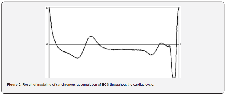
Application of the developed method of temporal scaling of quasi-periodic bioelectrical signals based on ultra-precise synchronization methods allows synchronous accumulation of ECS throughout the cardiac cycle. An example of modeling of such accumulation is shown in Figure 6.
Conclusion
The developed method makes it possible to study with high accuracy the interperiodic characteristics of electrocardiac signals throughout their cycle, including previously inaccessible areas outside the QRS-complex zone. This method is applicable for any quasi-periodic random processes. Development of new high-precision synchronization algorithms, new hardware solutions in the field of acquisition and recording of electrocardiac signals [4,6] and new methods of synchronous analysis of ECS SVR is carried out by the authors’ team in order to develop more effective approaches and techniques of secondary processing. This makes it possible to detect signs of pathological changes in the cardiovascular system [4], when classical methods do not give tangible results.
Acknowledgements
The present work was supported by Education and Science Ministry of Russian Federation, State task No. 075-00761-22-00, project No. FZZM-2022-0011.
Conflict of Interests
We declare that no conflict of interests exists.
References
- Zaichenko KV, Kordyukova AA, Sonin DL, Galagudza MM (2023) Ultra-high-resolution electrocardiography enables earlier detection of transmural and subendocardial myocardial ischemia compared to conventional electrocardiography. Diagnostics (Basel) 13(17): 2795.
- Aristarkhov GM, Gulyaev YV, Dmitriev VF, Komarov VV, Meshchanov VP, et al. (2024) Modern radio signals filtering devices. Methods, technologies, structures. Singapore: Bentham Science Publishing.
- Garvey JL (2006) ECG techniques and technologies. Emerg Med Clin N Am 24(1): 209-225.
- Zaichenko KV, Gurevich BS, Kordyukova AA (2021) Method of reliable electrocardiographic control of ischemia appearance in investigations with experimental animals. Proceedings - 2021 Ural Symposium on Biomedical Engineering, Radioelectronics and Information Technology, USBEREIT. Russia.
- Zaichenko KV, Afanasenko AS, Shevyakov DO (2024) Application of synchronous capture for ultra-high resolution electrocardiography. Biomed Eng 57: 350-352.
- Roth GA, Mensah GA, Johnson CO, Addolorato G, Ammirati E, et al. (2020) Global burden of cardiovascular diseases and risk factors, 1990-2019: Update from the GBD 2019 study. J Am Coll Cardiol 76(25): 2982-3021.


